
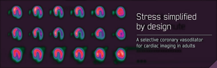
Trial design
Each patient received an initial stress scan using adenosine (6-minute infusion using a dose of 140 mcg/kg/min, without exercise) with a radionuclide gated SPECT (single photon emission computed tomography) imaging protocol. After the initial scan, patients were randomised (2 to 1) to either Rapiscan or adenosine, and received a second stress scan with the same radionuclide imaging protocol as that used for the initial scan (see figure)
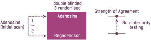
The primary objective of the studies was to demonstrate non-inferiority in the strength of agreement between sequential adenosine and Rapiscan images, and the strength of agreement between two sequential adenosine images for detecting the extent of reversible perfusion defects. Three independent expert readers who were blinded to treatment assignment performed image assessment.